
Industry Frontier | Pig kidney-derived cell-free DNA (xd-cfDNA) enables early warning of xenogeneic kidney transplant rejection
Recently, a study published in the journal Xenotransplantation provides a new and important direction for postoperative monitoring of kidney xenotransplantation. The study was completed by a team of Korean scientists. The results showed that by detecting xenogeneic cell-free DNA (xd-cfDNA) in the recipient's blood, traditional kidney function can be improved.
Recently, a study published in the journal Xenotransplantation provides a new and important direction for postoperative monitoring of kidney xenotransplantation. The study was completed by a team of Korean scientists. The results show that by detecting xenogeneic cell-free DNA (xd-cfDNA) in the recipient's blood, signs of damage to the transplanted kidney can be detected earlier and more sensitively before abnormalities in traditional kidney function indicators appear. In particular, it has outstanding early warning value for antibody-mediated rejection (AMR).
Research background
Kidney xenotransplantation is viewed as a potentially important way to address organ shortages. However, rejection after transplantation is a critical obstacle to long-term graft survival. At present, clinical monitoring mainly relies on renal function indicators such as serum creatinine (Cr) and blood urea nitrogen (BUN), but these indicators often become abnormal after significant damage to the transplanted kidney has occurred. Although kidney transplant biopsy can confirm the diagnosis, its invasive nature limits its frequent implementation. Therefore, it is crucial to develop a non-invasive, highly sensitive and early warning biomarker. The value of monitoring donor-derived cell-free DNA (dd-cfDNA) in allogeneic transplantation has been demonstrated, but the efficacy of this strategy in xenotransplantation has previously been unclear.
research methods
The research team transplanted minipig kidneys that had undergone multiple gene editing (knocking out multiple carbohydrate antigen genes that mediate hyperacute rejection and knocking in multiple human protective genes) into 10 cynomolgus monkeys. Plasma xd-cfDNA was collected and detected 1 week before surgery and 2, 4, 6, and 8 weeks after surgery or at the end point.
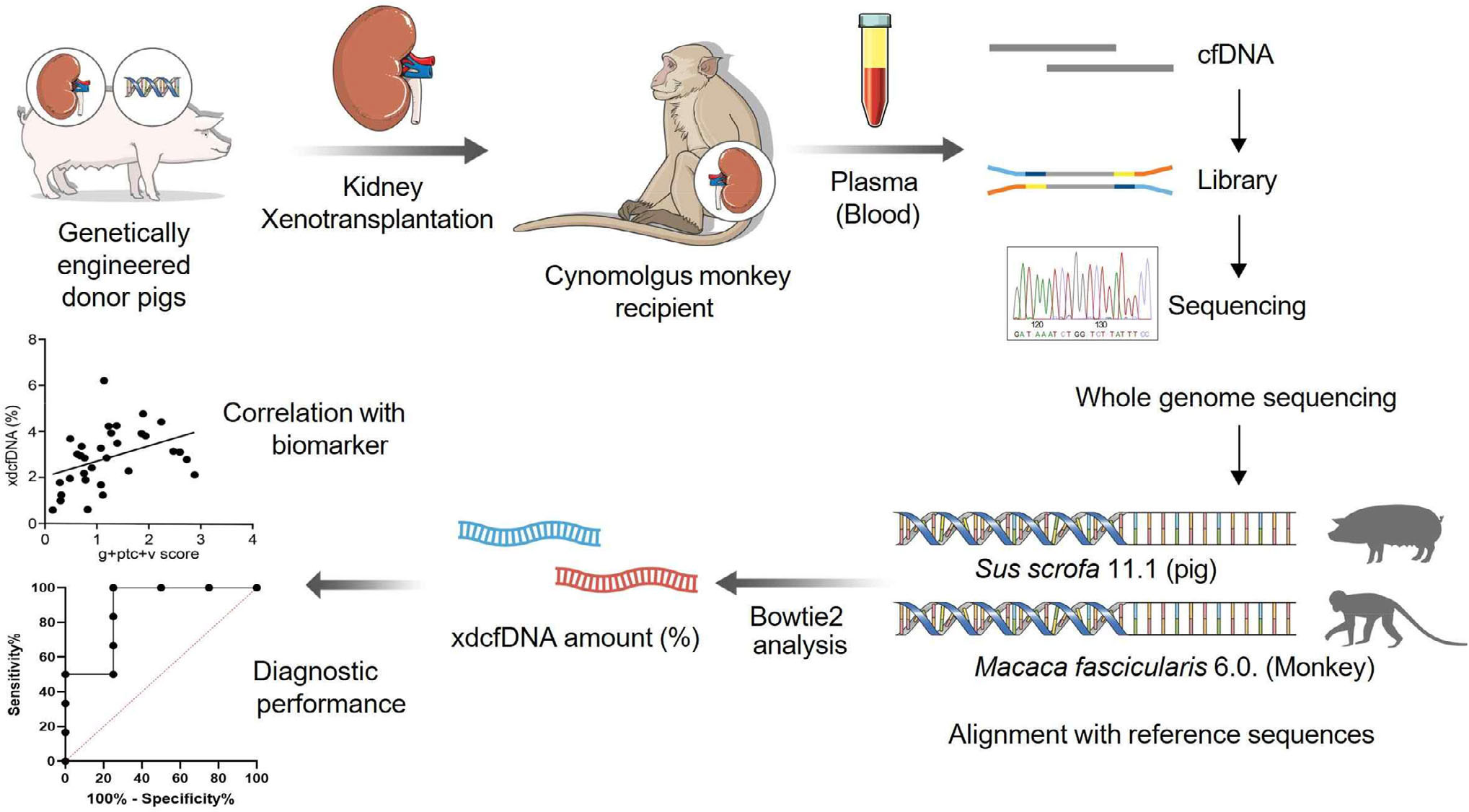
Figure 1. Flowchart of xenotransplantation experimental design
Core findings
01 Exclusion risk warning window advanced
Plasma xd-cfDNA levels began to rise 1 to 2 weeks before serum Cr and BUN significantly increased, achieving an earlier indication of the risk of rejection. (Figure 2)
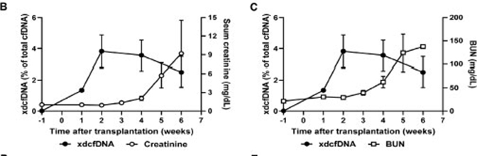
Figure 2. Comparison of changes in xd-cfDNA and renal function indicators
02 Excellent diagnostic performance
The study determined that the threshold value of plasma xd-cfDNA for diagnosing rejection was 2.545%. Based on the current study cohort, the sensitivity and specificity of xd-cfDNA in distinguishing "rejection" from "no rejection" at this threshold reached 100% (95% CI 64.57%–100% / 43.85%–100%) (Figure 3.). At the same time, the researchers proposed a more sensitive early warning threshold of 1.25% for early screening. When xd-cfDNA exceeds the warning threshold, it is prompted to strengthen monitoring and supplement it with other tests (such as donor-specific antibody testing); if its level continues to exceed the diagnostic threshold, transplant kidney biopsy is recommended to confirm the diagnosis, thereby guiding timely adjustment of the immunosuppression regimen.
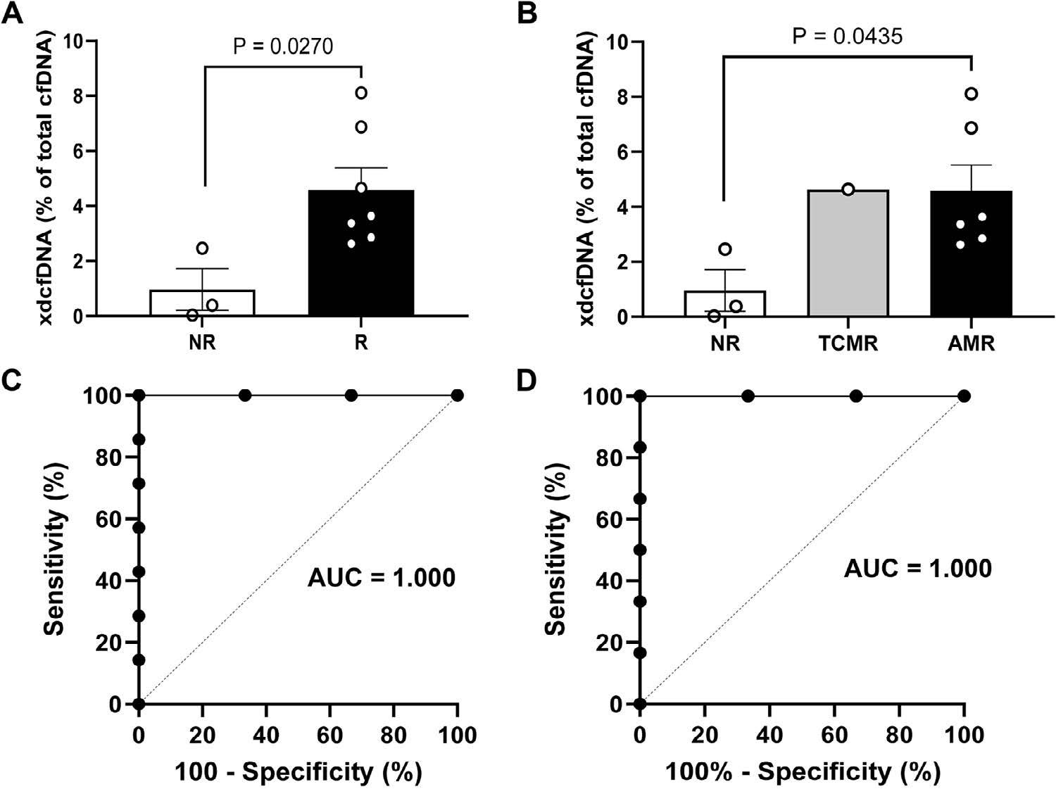
Figure 3. Association between xd-cfDNA and rejection 4 weeks after transplantation
A. Comparison of xd-cfDNA levels between rejection group and negative group; B. Comparison of xd-cfDNA levels between TCMR group, AMR group and negative group; C. ROC curve of xd-cfDNA for diagnosis of rejection; D. ROC curve of xd-cfDNA for diagnosis of AMR.
03 Closely associated with vascular damage
The level of xd-cfDNA is significantly correlated with vascular lesions (such as endovascular inflammation) and glomerular capillary IgG deposition in the Banff 2022 pathology scoring system, indicating that it can specifically reflect antibody-mediated vascular endothelial damage. (Figure 4)
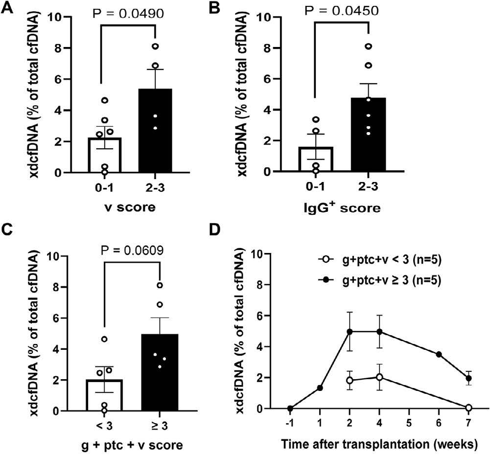
Figure 4. Correlation between xd-cfDNA levels and biopsy pathology scores
04 Indicates chronic injury process
In the group with chronic injury pathological changes such as renal tubular atrophy and interstitial fibrosis, the level of xd-cfDNA was also higher, suggesting that it may be useful in monitoring the chronic injury process of transplanted kidneys in the future. (Figure 5)
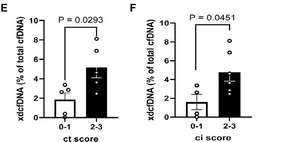
Figure 5. Comparison of xd-cfDNA in tubular atrophy subgroup and interstitial fibrosis subgroup
Summary
In recent years, clinical research on xenotransplantation has continued to advance, but long-term graft survival still faces challenges. Rejection may be one of the factors affecting long-term survival. How to detect rejection early and adjust immunosuppression regimens has become one of the research hotspots in the field of xenotransplantation. This study used xd-cfDNA derived from pig kidneys to detect rejection after transplantation, suggesting the great potential of xd-cfDNA as an early diagnostic biomarker for xenograft rejection. As the first domestic institution with independent intellectual property rights for dd-cfDNA and achieving widespread clinical application, AlloDx has also established a detection technology with independent intellectual property rights in the detection of xenogeneic cell-free DNA (xd-cfDNA) (a molecular marker and kit for monitoring rejection of pig xenografts, CN119552954A, December 28, 2023). It can accurately quantify xd-cfDNA after xenotransplantation and is expected to provide a powerful tool for clinical research on xenotransplantation.
Reference sources:
Han K-H, et al. Xenograft-derived cell-free DNA as an early biomarker of rejection in genetically engineered pig-to-nonhuman primate kidney xenotransplantation.Xenotransplantation.2025;32:e70100.doi:10.1111/xen.70100
Some original figures, videos and downloadable materials are provided in Chinese.